
Developed by Albert Einstein College of Medicine and the Salk Institute for Biological Studies, Multicolor Imaging Technology Lets Researchers Track Proteins and Cellular Activity with Exceptional Precision and Minimal Background
, /PRNewswire/ — Fluorescent probes have transformed modern biology by allowing researchers to tag and visualize individual molecules in living cells, tissues, and animals. Using these tools, researchers can watch viruses infect cells in real time, observe cellular trash collection, and track the signaling that spurs tumor growth. Now, scientists at Albert Einstein College of Medicine and the Salk Institute for Biological Studies have developed a new molecular imaging technology that illuminates proteins inside living cells and animals far more clearly than before. Described in today’s issue of Nature Methods, the system uses engineered fluorescent nanobodies—tiny antibody-like protein fragments—that light up only when they bind to their specific targets.

“The key advantage of our approach is that the signal appears only where the target protein is present,” said Vladislav Verkhusha, Ph.D., a co-corresponding author on the study and professor of genetics and co-director of the Gruss-Lipper Biophotonics Center at Einstein. “That eliminates the background glow that has long limited the precision of intracellular imaging.” The study’s other co-corresponding author is Axel Nimmerjahn, Ph.D., professor at Salk.
Solving a Key Imaging Problem
Over the past decade, fluorescent nanobodies have emerged as powerful tools because they bind to specific proteins in living cells. However, conventional versions glow whether or not they are attached to their targets, producing diffuse background signals that obscure fine details.
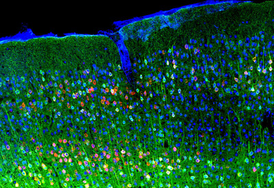
To overcome this limitation, the researchers engineered a new class of probes called VIS-Fbs (visible-spectrum target-stabilizable fluorescent nanobodies). These probes rapidly degrade if they do not bind to their intended target; only when bound do they become stable and brightly fluorescent. This “on-demand” fluorescence reduces background noise by as much as 100-fold, enabling much sharper visualization of protein location and dynamics.
The researchers created versions of their VIS-Fb probes that fluoresce across nearly the entire visible spectrum, from blue to far red, making it possible to track multiple proteins or cellular processes within the same living cell at once.
A Versatile Platform
Rather than create a single probe, Dr. Verkhusha and colleagues developed a modular engineering platform for building VIS-Fb probes that can be adapted to many targets and experimental needs. By integrating more than 20 different fluorescent proteins and biosensors into multiple nanobody scaffolds, they created a flexible toolkit with multiple capabilities.
With this approach, multiple proteins can be tracked simultaneously in different cellular compartments using VIS-Fb probes that emit distinct colors, enabling true multicolor imaging within the same cell. Certain VIS-Fb variants can also be activated, or switched on and off, with light, making it possible to follow protein behavior over time with high spatial and temporal precision. Incorporating biosensors for ions and metabolites further allows the probes to report not only where proteins are located but also what they are doing in real time, providing direct insight into cellular activity. In addition, combining stable reference signals with activity-sensitive fluorescence enables ratiometric measurements that improve the accuracy of quantifying cellular processes, even in complex environments such as living brain tissue.
“The VIS-Fb approach allows us to identify and track specific cell populations in living organisms based on the proteins they express, rather than just their location,” said Natalia Barykina, Ph.D., the first author of the study and a postdoctoral fellow in Dr. Verkhusha’s lab.
The researchers demonstrated the system in a range of living models. In mice, VIS-Fb probes enabled precise imaging of central nervous system activity in neurons and astrocytes, with strong signal quality during behavior. In zebrafish embryos, the technology allowed real-time tracking of dynamic changes during early development and in response to drugs that alter signaling pathways.
“Our results show that this imaging platform offers a much clearer and more precise view of how proteins behave inside living systems,” Dr. Verkhusha said. “It opens the door to studying complex biological processes, such as cell signaling, development, and disease progression, in new ways.”
Additional Einstein authors include Juliana Mendoça-Gomes, Ph.D. and Sofia de Oliveira, Ph.D. Additional authors are Erin Carey, Salk Institute for Biological Studies, and Olena Oliinyk, Ph.D., University of Helsinki, Finland.
The title of the paper is “Synthetic multicolor antigen-stabilizable nanobody platform for intersectional labelling and functional imaging.” Funding sources include the National Institutes of Health: GM122567 (V.V.), NS123719 (A.N.), and GM118027 (S.dO); the Jane and Aatos Erkko Foundation (220011); the Research Council of Finland (360277); the Chan Zuckerberg Initiative Foundation (MET-000000045); the NOMIS Foundation Neuroimmunology Initiative; and the Edwards-Yeckel Research Foundation.
About Albert Einstein College of Medicine
Albert Einstein College of Medicine is one of the nation’s premier academic centers for basic science research, clinical investigation, and biomedical education. Located in the Bronx, Einstein is home to nearly 1,000 M.D., Ph.D., and M.D./Ph.D. students and more than 2,000 full-time faculty members. Einstein receives approximately $200M in funding from the National Institutes of Health (NIH) each year and houses six NIH-funded research centers, in cancer, intellectual and developmental disabilities, clinical and translational research, AIDS, and two in diabetes. In partnership with Montefiore Health System, Einstein advances clinical and translational research to accelerate the pace at which new discoveries become the treatments that benefit patients. For more information, please visit einsteinmed.edu, and follow us on Instagram, LinkedIn, Twitter, Facebook, and view us on YouTube.
SOURCE Albert Einstein College of Medicine


